Prevention of the disease caused by Actinobacillus pleuropneumoniae

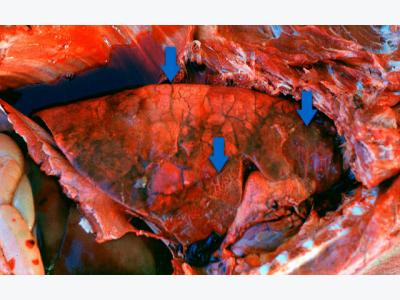
Acute pleuropneumonia in swine: three observations (indicated by arrows) can frequently be made: consolidated areas that are from dark red to black, interlobular edema and fibrinouspleuritis. Picture courtesy of Dr Robert Desrosiers
This article explains the main preventive measures against App, focusing on the use of antimicrobials and vaccines.
Once clinical disease of pleuropneumonia has been confirmed through laboratory examination, prevention measures must be applied.
Antimicrobials: If the appearance of clinical signs is predictable, antimicrobial treatments before specific high risk periods can be established. The reinforcement of political restrictions on the use of antimicrobials in order to reduce antimicrobial resistance in veterinary medicine makes this practice difficult/impossible in some European countries. However, the prophylactic/metaphylactic prevention of swine pleuropneumonia is still commonly used in other European as well as most American and Asian countries. There is not a clear recommendation about which specific antimicrobial drugs must be used: it is important to isolate the App involved in the case and to perform an antibiogram to correctly choose the antimicrobial. It has been reported that the massive use of very strong bactericidal antimicrobials may prevent the development of immunity in treated animals: once the antimicrobial treatment is stopped, clinical cases come back (bacteria remain hidden in the tonsils). The next question is: how much time the treatment must be kept? Usually, treatments should be kept for 2-3 weeks, but sometimes longer periods of time are needed to prevent new clinical cases. When disease appears in finisher animals, antimicrobials with shorter withdrawal periods must be chosen. This kind of prevention measures should be temporary. The use of vaccination is recommended to reduce antimicrobial use. However, under extreme conditions (highly virulent strains), antimicrobial support might be necessary to complete protection given by vaccines. Antimicrobial treatments do not eliminate App from tonsils of carrier animals.
Vaccination: There are different types of commercial vaccines available. These vaccines can be classified as:
1. Bacterins (washed and killed whole bacteria): Protection given by this type of vaccine is serotype-specific: you need to know which serotype is involved in the farm. It is unusual (not impossible when different origins are mixed) to have more than one serotype causing disease in a single farm: a good diagnosis is needed. Antibodies raised against this vaccine are directed to "bacterial body antigens": capsule, surface proteins, bacterial cell wall, etc. When these antibodies are present in the animal lung, they "attach" to bacteria and alveolar macrophages and other cells will ingest and destroy App. In the absence of these antibodies, phagocytic cells cannot “capture” App bacteria, which reproduce and produce, among other products, the toxins and cause lesions.
2. Purified toxoid-based vaccines (sometimes enriched with surface proteins): ApxI, ApxII and ApxIII toxoids are present. Different strains of App produce one or two of these toxins. Protection given by this type of vaccine is usually against all serotypes, since all App strains produce one or two of the toxins included. Antibodies raised against this vaccine "neutralize" the toxins: just prevent them to cause lesions. Antibodies do not react with App bacteria: toxins are secreted. So, App can still reproduce in the lungs, although toxic effect of toxins is neutralized
3. "Mixed": Bacterin (for specific serotypes) + purified toxoid-based vaccine: it is a combination of "1" and "2". Antibodies produced are anti-toxins and anti-bacterial components (only for serotypes included in the vaccine).
Please note that autogenous vaccines are usually "bacterins". Its use should be restricted only to cases when the decision of using a bacterin has been taken and the serotype involved is either unknown or absent from commercial vaccines. Strains belonging to a given serotype are antigenically very similar around the world: a commercial vaccine produced with a serotype 2 isolated in Europe will protect against a serotype 2 isolated in Asia.

Acute pleuropneumonia in swine: three observations (indicated by arrows) can frequently be made: consolidated areas that are from dark red to black, interlobular edema and, in this picture, subtle fibrinouspleuritis. Picture courtesy of Dr Robert
Vaccination of piglets
• Strong interference with maternal antibodies: usually, first dose should not be applied before 7-8 weeks of age.
• Never use a single dose: a booster reaction is needed.
• Avoid vaccination when high level of transmission of PRRS virus occurs at the same time among piglets.
• Adjust age of vaccination if needed, depending on the age when clinical signs appear, especially those that occur at the end of the finishing period: some commercial vaccines have the information of late protection. Otherwise, the vaccination can be moved later to keep high protection at the moment of the presence of clinical signs. Please allow at least 5-6 weeks after the first dose to reach good protection.
Vaccination of sows/gilts
• Two doses (second dose 3 weeks before farrowing) the first time; repeat one dose 3 weeks before farrowing each time
• Increase the level of maternal antibodies when clinical cases are present in young animals (not very usual).
• Increase the level of maternal antibodies that reduce/delay colonization of piglets: in multi-site production system, these maternal antibodies would reduce the prevalence of carrier animals at weaning (around 3 weeks of age). Reduction of prevalence may reduce/eliminate the presence of clinical signs in grower-finisher animals. Only bacterins (with antibodies against the whole bacteria) can induce such reduction of colonization: antibodies "attached" to the whole bacteria will prevent/reduce App colonization of tonsils. Antibodies against secreted toxins (toxoid-based vaccines) do not have such effect.
• Vaccination of replacement gilts during quarantine can be done. If the objective is to protect App free highly susceptible animals (prevent disease), vaccination with either a bacterin or a toxoid-based vaccine can be done.
• High level of antibodies (after infection or vaccination) do not eliminate App from tonsils of carrier animals.
How to evaluate antibody response in vaccinated animals?
• If a bacterin is used: None of the commercial kits available (LPS-based ELISA tests) are able to accurately evaluate response to vaccination. These tests have been standardized to detect infection, not vaccination.
• Antibodies induced against vaccination are mainly those directed to the bacterial capsule, proteins and some of them against the LPS. The antigen used in those ELISA test is a purified LPS: it is possible that most antibodies produced after vaccination do not react with this antigen: A NEGATIVE ELISA TEST AFTER VACCINATION DOES NOT IMPLY LACK OF ANTIBODY RESPONSE AGAINST THE VACCINE.
• Even if antibodies against the LPS may be produced after vaccination, the commercial kits available have been standardized to detect levels of antibodies after infection, not vaccination. So, dilutions of sera needed to be done to carry out the test may induce negative results when evaluating antibody response against vaccination.
• Only customised ELISA tests, using whole bacteria as antigen (as that used in the vaccine) can be used to measure antibody response. Antibodies titers before and after vaccination can then be compared.
• If a toxin-based vaccine is used: there is no commercial test to measure antibodies against ApxI, II and III. These tests can only be done by vaccine-producing companies since purified antigens are needed. The ApxIV ELISA test cannot be used, since this toxin is never produced after vaccination (infection only).
Other measures to increase prevetion of App disease
• Strict use of "all in-all out" systems
• Use of batch farrowing
• Control of the environment (temperature, ventilation, etc.)
• Control of predisposal infections: mainly SIV and Mycoplasma hyopneumoniae.
Có thể bạn quan tâm
Phần mềm

Phối trộn thức ăn chăn nuôi

Pha dung dịch thủy canh

Định mức cho tôm ăn

Phối trộn phân bón NPK

Xác định tỷ lệ tôm sống

Chuyển đổi đơn vị phân bón

Xác định công suất sục khí

Chuyển đổi đơn vị tôm

Tính diện tích nhà kính

Tính thể tích ao hồ


 Insects as an alternative raw material in pig…
  Benefits of corn co-products from wet milling for…